1,640 neurons imaged: New 3.5-gram MiniXL microscope lets scientists watch multiple brain regions at once
For years, scientists studying the neural basis of behavior in mice faced a difficult trade-off. Existing miniature microscopes either had a very small field of view, limiting them to observing a single small patch of brain tissue, or were too heavy for the mice to behave naturally. This made it incredibly difficult to study how different brain regions coordinate to produce complex behaviors like social interaction or memory formation.
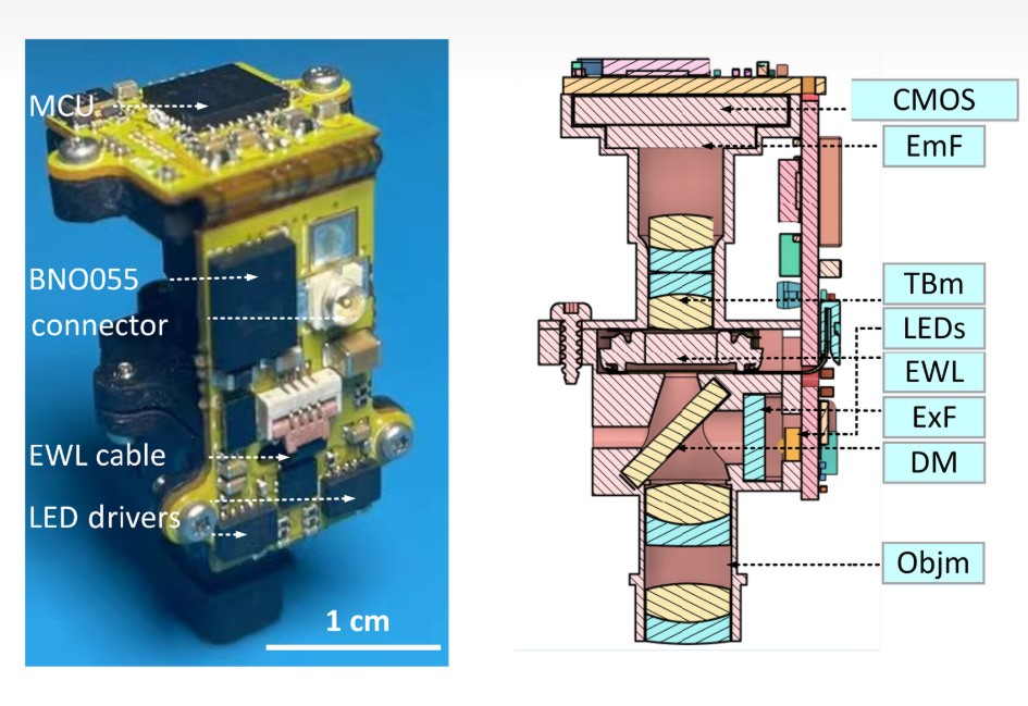
Now, the Miniscope extra Large FOV (MiniXL), a 3.5 g head-mounted miniscope, is pushing the boundaries of neuroscience after successfully imaging 1,640 neurons across a 3.5-mm-diameter field-of-view (FOV) — 9x larger than its predecessor’s 1 mm². It tracked 44–53% place cells in hippocampal CA1 on a 2 m linear track, imaged left and right medial prefrontal cortex (mPFC), and mPFC with nucleus accumbens (NAc) during social interactions in mice, using a 5-megapixel CMOS sensor and a single 0.3 mm coaxial cable for data streaming. This achievement was announced on June 11, 2025.
Its predecessors: Miniscope V4 and MiniLFOV, were either too heavy or too weak. MiniLFOV was capable of imaging ~2,000 neurons at once, but it weighed 13.9 g. The Miniscope V4 weighed 2.6 g but was only capable of imaging ~200 neurons at once. One could say that the Miniscope extra Large FOV (MiniXL) played its role as the Immanuel Kant of the Miniscope world by successfully reconciling the strengths and weaknesses of its predecessors.
Here is how it compares to its predecessors (Miniscope V4 and MiniLFOV):
| Feature | MiniXL (New) | Miniscope V4 | MiniLFOV |
|---|---|---|---|
| Weight (g) | 3.5 | 2.6 | 13.9 |
| FOV | approximately 10 mm² | 1 mm² | 9.72 mm² |
| Imaged neurons | 1640 across 3 sessions | ~200 | ~2000 |
| Min. animal (size) | Mice | Mice | Rats |
Its dual-region imaging capability, highlighted here by simultaneous imaging of the mPFC and NAc, addresses a critical need for tools that can explore the neural basis of behaviors coordinated by multiple brain regions — UCLA research team.
Beyond its technical specifications, another interesting aspect of the MiniXL is its open-source design. As part of the UCLA Miniscope project, all design files and documentation are being made freely available online, aiming to lower technical and economic barriers and accelerate neuroscience research globally.
While this technology is a leap for laboratory research, its application in human medicine is still a distant prospect.
Source(s)
Science Advances and LABmaker: Miniscope V4 and MiniLFOV
